Free Online Productivity Tools
i2Speak
i2Symbol
i2OCR
iTex2Img
iWeb2Print
iWeb2Shot
i2Type
iPdf2Split
iPdf2Merge
i2Bopomofo
i2Arabic
i2Style
i2Image
i2PDF
iLatex2Rtf
Sci2ools
ISBI
2006
IEEE
2006
IEEE
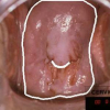
Automatic landmark detection in uterine cervix images for indexing in a content-retrieval system
This work is motivated by the need for visual information extraction and management in the growing field of content based image retrieval from medical archives. In particular it focuses on a unique medical repository of cervicographic images ("Cervigrams") collected by the National Cancer Institute, National Institutes of Health, to study the evolution of lesions related to cervical cancer. The paper briefly presents a framework for cervigram segmentation and labelling, focusing on the identification of two anatomical landmarks: the cervix boundary and the os. These landmarks are identified based on their convexity, using adequate mathematical tools. Segmentation results are exemplified and an initial validation is carried out on a subset of 120 manually labelled cervigrams.
Adequate Mathematical Tools | ISBI 2006 | Medical Imaging | National Cancer Institute | Unique Medical Repository |
| Added | 20 Nov 2009 |
| Updated | 20 Nov 2009 |
| Type | Conference |
| Year | 2006 |
| Where | ISBI |
| Authors | Gali Zimmerman, Shiri Gordon, Hayit Greenspan |
Comments (0)

