Free Online Productivity Tools
i2Speak
i2Symbol
i2OCR
iTex2Img
iWeb2Print
iWeb2Shot
i2Type
iPdf2Split
iPdf2Merge
i2Bopomofo
i2Arabic
i2Style
i2Image
i2PDF
iLatex2Rtf
Sci2ools
142
click to vote
ISBI
2002
IEEE
2002
IEEE
Statistical shape models for segmentation and structural analysis
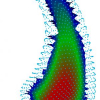
Biomedical imaging of large patient populations, both cross-sectionally and longitudinally, is becoming a standard technique for noninvasive, in-vivo studies of the pathophysiology of diseases and for monitoring drug treatment. In radiation oncology, imaging and extraction of anatomical organ geometry is a routine procedure for therapy planning an monitoring, and similar procedures are vital for surgical planning and image-guided therapy. Bottlenecks of today's studies, often processed by labor-intensive manual region drawing, are the lack of efficient, reliable tools for threedimensional organ segmentation and for advanced morphologic characterization. This paper discusses current research and development focused towards building of statistical shape models, used for automatic model-based segmentation and for shape analysis and discrimination. We build statistical shape models which describe the geometric variability and image intensity characteristics of anatomical structures. ...
ISBI 2002 | Medical Imaging | Shape Analysis | Statistical Shape Models | Threedimensional Organ Segmentation |
Related Content
| Added | 20 Nov 2009 |
| Updated | 20 Nov 2009 |
| Type | Conference |
| Year | 2002 |
| Where | ISBI |
| Authors | Guido Gerig, Martin Andreas Styner, Gábor Székely |
Comments (0)

