Free Online Productivity Tools
i2Speak
i2Symbol
i2OCR
iTex2Img
iWeb2Print
iWeb2Shot
i2Type
iPdf2Split
iPdf2Merge
i2Bopomofo
i2Arabic
i2Style
i2Image
i2PDF
iLatex2Rtf
Sci2ools
ISBI
2006
IEEE
2006
IEEE
Mapping ventricular changes related to dementia and mild cognitive impairment in a large community-based cohort
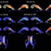
We present a fully-automated technique for visualizing localized cerebral ventricle shape differences between large clinical subject groups who have received a magnetic resonance (MR) image scan. The technique combines a robust, automated technique for ventricular segmentation with a 3D surfacebased radial thickness mapping approach that allows spatiallylocalized statistical tests of relative shape differences between clinical groups. The technique is used to analyze localized ventricular expansion in Alzheimer's Disease (AD) and mild cognitive impairment (MCI) in a large cohort of communitydwelling elderly individuals (N=339). The resulting maps are the first to chart localized ventricular dilation in a cohort of this size. Besides showing patterns of ventricular expansion that may be consistent with the spatial progression of ADrelated pathology, the maps reveal new information about localized ventricular atrophy that may have been overlooked to date. A detailed understanding o...
ISBI 2006 | Localized Ventricular Dilation | Localized Ventricular Expansion | Medical Imaging | Ventricular Atrophy |
| Added | 20 Nov 2009 |
| Updated | 20 Nov 2009 |
| Type | Conference |
| Year | 2006 |
| Where | ISBI |
| Authors | Owen T. Carmichael, Paul M. Thompson, Rebecca A. Dutton, Allen Lu, Sharon E. Lee, Jessica Y. Lee, Lewis H. Kuller, Oscar L. Lopez, Howard Aizenstein, Carolyn C. Meltzer, Yanxi Liu, Arthur W. Toga, James T. Becker |
Comments (0)

