Free Online Productivity Tools
i2Speak
i2Symbol
i2OCR
iTex2Img
iWeb2Print
iWeb2Shot
i2Type
iPdf2Split
iPdf2Merge
i2Bopomofo
i2Arabic
i2Style
i2Image
i2PDF
iLatex2Rtf
Sci2ools
113
click to vote
ISBI
2008
IEEE
2008
IEEE
A quantification framework for post-lesion neo-vascularization in retinal angiography
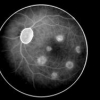
We describe an image processing framework designed to detect and quantify the genesis of microscopic choroidal blood vessels. We used fluorescein angiography to monitor the dynamic of neo-vascularization of the retina after inducing lesions with a calibrated laser pulse. The angiogenesis can be revealed by an increase in the overall fluorescence level and/or diffusion size of the lesion. The proposed framework allows measuring both features from mis-aligned angiograms acquired with different gains and contrasts. It consists in aligning all the images, homogeneizing their intensity characteristics and segmenting the lesions. In particular, we implemented a level set segmentation algorithm to delineate the diffusion area. We show that our framework allows detecting neo-veovascularization when one of these features changes by less than 10%.
Image Processing Framework | ISBI 2008 | Level And/or Diffusion | Medical Imaging | Microscopic Choroidal Blood |
| Added | 20 Nov 2009 |
| Updated | 20 Nov 2009 |
| Type | Conference |
| Year | 2008 |
| Where | ISBI |
| Authors | Sylvain Takerkart, Romain Fenouil, Jérome Piovano, Alexandre Reynaud, Louis Hoffart, Frederic Chavane, Théodore Papadopoulo, John Conrath, Guillaume S. Masson |
Comments (0)

