ISBI
2008
IEEE
15 years 4 days ago
2008
IEEE
ISBI
2008
IEEE
15 years 4 days ago
2008
IEEE
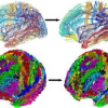
This work exploits the idea that each individual brain region has a specific connection profile to create parcellations of the cortical surface using MR diffusion imaging. The par...
ISBI
2008
IEEE
15 years 4 days ago
2008
IEEE
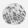
We present a novel use of GPUs (Graphics Processing Units) for the analysis of histopathologicalimages of neuroblastoma, a childhood cancer. Thanks to the advent of modern microsc...
ISBI
2008
IEEE
15 years 4 days ago
2008
IEEE
Reconstruction algorithms for Optical Diffuse Tomography (ODT) rely heavily on fast and accurate forward models. Arbitrary geometries and boundary conditions need to be handled ri...
ISBI
2008
IEEE
15 years 4 days ago
2008
IEEE
Model-based segmentation approaches, such as those employing Active Shape Models (ASMs), have proved to be useful for medical image segmentation and understanding. To build the mo...
ISBI
2008
IEEE
15 years 4 days ago
2008
IEEE
We propose a quantified asymmetry based method for age estimation. Our method uses machine learning to discover automatically the most discriminative asymmetry feature set from di...
ISBI
2008
IEEE
15 years 4 days ago
2008
IEEE
We propose a novel automatic method to segment the myocardium on late-enhancement cardiac MR (LE CMR) images with a multi-step approach. First, in each slice of the LE CMR volume,...
ISBI
2008
IEEE
15 years 4 days ago
2008
IEEE
Neuroimaging at the group level requires spatial normalization across individuals. This issue has been receiving considerable attention from multiple research groups. Here we sugg...
ISBI
2008
IEEE
15 years 4 days ago
2008
IEEE
GRAPPA is one of the predominant methods used to reconstruct accelerated parallel MRI data. In has been shown previously that spatially varying the GRAPPA reconstruction coefficie...
ISBI
2008
IEEE
15 years 4 days ago
2008
IEEE
In this paper, we adress the question of decoding cognitive information from functional Magnetic Resonance (MR) images using classification techniques. The main bottleneck for acc...


